

Scoliosis Surgery Procedure Prices and Aftercare in Turkey
- What is scoliosis?
- What forms of scoliosis are there?
- Frequency: How often the disease occurs
- How is scoliosis treated?
- Scoliosis Surgery
- Rehabilitation
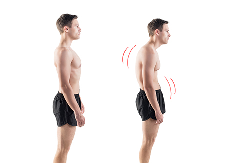
- Scoliosis symptoms
- Scoliosis causes and risk factors
- Diagnosis of Scoliosis
- Course and prognosis of Scoliosis
- Complications after scoliosis surgery
- Prevention
In scoliosis, the spine curves sideways. The vertebral bodies are usually twisted as well. Symptoms usually only appear with a stronger curvature of the spine. Mild forms can often be treated with physiotherapy and a special corset, while severe cases require surgery.
- Treatment: physiotherapy, corset, cast, clamps, surgery, special exercises
- Symptoms: shoulders at different heights, crooked pelvis, crooked head, lateral "rib hump", back pain, tension
- Causes and risk factors: predominantly unknown cause; secondary scoliosis caused by congenital or acquired diseases or injuries
- Diagnosis: Physical examination, Adams test, flexibility/strength tests, X-ray, assessment of skeletal maturity
- Prognosis: Usually good prognosis with treatment; the earlier the therapy, the better the prognosis; untreated Progression of the disease, stiffening of the respective vertebral section, early wear and tear
- Prevention: Concrete prevention is usually not possible; Early detection and therapy prevent later consequences
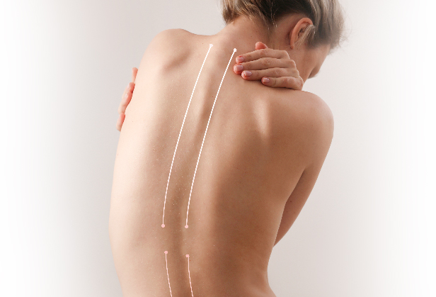
What is scoliosis?
Scoliosis is a permanent lateral curvature of the spine , in which the vertebral bodies themselves are also twisted and misaligned. In order to understand exactly what constitutes scoliosis, it is helpful to know how a healthy spine is structured.
Short excursion into anatomy: The structure of the spine
A healthy spine consists of about 33 vertebrae: seven cervical vertebrae, twelve thoracic vertebrae, five lumbar vertebrae, five fused sacral vertebrae, and about four — also fused — coccygeal vertebrae. The vertebral bodies are connected to the neighboring vertebrae and ribs via bone processes.Viewed from the side, the spine is shaped like a double "S". The cervical and lumbar spine each curve forward ( lordosis ), the thoracic and sacral spine ( sacrum ) backward ( kyphosis ). If you look at the spine from behind, it forms an almost straight line with its spinous processes from the head to the anal fold. The vertebral bodies lie evenly on top of each other and between each two of them there is an intervertebral disc as a shock absorber.
The spine is an important part of the supporting skeleton and also protects the spinal cord, a bundle of nerve pathways that carries signals between the body and the brain .
The scoliosis
In the case of scoliosis, the structure of the spine is disturbed. The name of the disease derives from the Greek word "skolios", which means "crooked": In this case, the spine not only curves forwards and backwards, but also sideways.According to the guideline-based definition of scoliosis, this clinical picture is "a permanently existing (fixed) lateral spinal curvature of at least ten degrees Cobb angle ". This angle indicates how strong the lateral curvature of the spine is and can be determined using an X-ray image. Depending on which side the spine curves to, doctors speak of a right or left convex scoliosis.
In addition, the individual vertebrae and the entire spine are twisted in their longitudinal axis (rotation and torsion). As a result, the bony vertebral body processes ( spinous process , processus spinosus) do not point straight backwards. So the side of the processes that faces the abdomen or chest rotates in the direction of the curvature of the spine. The rotation is strongest at the apex of the scoliosis and decreases again at the foothills of the curved section of the spine.
As the scoliosis progresses, it is possible for the corresponding section of the vertebra to stiffen.
Due to the different degrees of twisting, tension and pressure forces arise between the individual vertebral bodies. As a result, the vertebral bone also has a distorted bone structure (torqued): the vertebral body is higher on the side that curves outwards than on the side that points inwards. The same applies to the intervertebral discs between the vertebrae. This results in a permanently existing crooked growth. Experts also refer to the twisted and crooked spine as torsional scoliosis .
Torsion scoliosis usually only occurs on the main curve. In order to compensate for severe scoliosis, muscle power creates secondary curves in the spine in the immediate vicinity of the main curve (static compensation). However, the minor curves do not exhibit any rotation or torsion. If it does, it's called multiple scoliosis .
What forms of scoliosis are there?
Depending on the point of view, scoliosis can be divided into different forms. For example, a general distinction is made between idiopathic scoliosis and secondary scoliosis .
- Idiopathic means that no specific trigger for the disease can be found.
- Secondary scoliosis, on the other hand, is always the result of a known cause.
A scoliotic malposition passes and normalizes itself again through passive or active movements. It is created, for example, to compensate for a pelvic obliquity.
Since in many cases the cause of scoliosis is not known, it cannot be effectively prevented.
Real scoliosis can be further differentiated according to age and curvature pattern.
Scoliosis of different age groups
Among other things, scoliosis can also be differentiated according to the time at which it first appeared. Doctors call the early form infant scoliosis . In most cases, it resolves without treatment. Doctors speak of infantile scoliosis when the curvature of the spine occurs by the age of three. Scoliosis in children between the ages of four and ten is referred to as the juvenile form .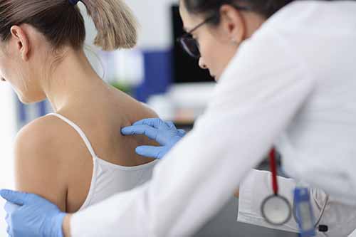
However, adolescent scoliosis is most common from the age of eleven. The spine is usually curved to the right in the thoracic vertebrae area (right-convex scoliosis). Girls are affected more often than boys.
Curvature pattern
In addition, a scoliosis can be assigned to the middle (or the vertex) of its main curvature in the spine. In a thoracic scoliosis , the curvature is in the area of the thoracic spine (thoracic spine). Thoraco-lumbar scoliosis has its strongest lateral deflection where the thoracic spine merges into the lumbar spine (lumbar spine). A curvature of the spine in the lumbar area is called lumbar scoliosis .
- In some cases, those affected suffer from thoracic and lumbar scoliosis at the same time. A curvature pattern is formed which - when looking at the patient's back from behind - is reminiscent of the letter "S" (double arched).
- If the spine is completely bent to one side, doctors speak of a C-shaped scoliosis.
- If the spine curves in all sections (thoracic, lumbar and their transition) alternately to the right and left, a double S spine develops, also known as triple scoliosis.
Degree of curvature
Depending on how severely the spine is curved, scoliosis can be divided into:
- Mild scoliosis: angle up to 40 degrees (1st degree scoliosis)
- Moderate scoliosis: angle between 40 and 60 degrees (2nd degree scoliosis)
- Severe scoliosis: angle from 61 to 80 degrees (3rd degree scoliosis)
- Very severe scoliosis: angle greater than 80 degrees (4th degree scoliosis)
Frequency: How often the disease occurs
About two to five percent of the population suffer from idiopathic scoliosis. According to a study by the Maimonides Medical Center (USA), the frequency increases in old age (60 to 90 years) to up to 68 percent.The greater the curvature of the spine and the higher the age, the more often women or girls are affected. In boys, slight scoliosis can be observed. More severe scoliosis, with a Cobb angle greater than twenty degrees, is about seven times more common in women than in men.
Severe disability
In individual cases it is possible that scoliosis is a possible reason for a recognized degree of disability. However, this depends on the severity and the limitations of the disease for the individual. It is therefore possible for a severe scoliosis with a Cobb grade of 70 degrees or more to be recognized with a degree of disability (GdB) in the sense of a severe disability of 50 to 70 percent, with more severe limitations even more.The local pension offices are usually responsible for the recognition of a GdB; your doctor is the contact person.
How is scoliosis treated?
Physicians treat scoliosis conservatively with physiotherapy or corsets and, in severe cases, surgically. It is advisable to start scoliosis therapy as soon as possible after diagnosis. The choice of treatment depends on the extent, cause and location of the curvature of the spine, as well as the age and physical condition of the patient. In the case of mild scoliosis, physiotherapy is often sufficient, while doctors treat more severe forms with a scoliosis corset . If there is a very strong curvature, an operation often helps.
Goals of scoliosis therapy
When treating a curvature of the spine, doctors, together with other specialists such as physiotherapists, try to ensure that the scoliosis regresses or at least does not get worse.If the curvature can be reduced by scoliosis therapy, further treatment steps ensure that this success is maintained. The guidelines set a clear goal for children and adolescents: the Cobb angle should be less than 40 degrees at the end of growth. If this is successful, according to experts, surgical scoliosis therapy is no longer necessary.
Scoliosis Corset
A scoliosis corset is used if the child has severe curvatures of the spine (Cobb angle 20-50 degrees). Very good results are often achieved with scoliosis that is not due to serious underlying diseases (malformations, muscle or nerve diseases or others).The corset ( orthesis ) is made of plastic and has built-in pressure pads (pads) as well as free spaces (expansion zones).
It is custom-made, attached to the body with straps and Velcro fasteners and has the task of returning the spine to its natural shape. The patient usually wears the orthosis for 22 to 23 hours a day. Depending on the height of the main curves, different scoliosis corsets are available.
For girls, the daily wearing time can be gradually reduced, depending on the course, about two to three years after the first menstrual period. Boys should first have reached a certain skeletal maturity (Risser stage four or five), so that the spine is no longer expected to grow much.
Adults benefit little from this scoliosis therapy because their bone growth has already ended. Nevertheless, the orthoses are also used in old age, for example to stabilize and thus alleviate the course of the disease.
Regular gymnastic exercises also support successful scoliosis therapy with orthoses.
Plaster treatment
In some cases of early curvature of the spine (under the age of five, early-onset scoliosis), scoliosis therapy using a plaster corset can be considered. The spine continues to grow normally. The cast treatment is usually followed by therapy with a scoliosis corset.
Scoliosis Surgery
In some cases, conservative scoliosis therapy (physiotherapy, corset) is not sufficient. If a scoliosis is getting worse and the curvature is severe, doctors usually recommend surgical scoliosis therapy. In doing so, they take several factors into account:
- the severity of the curvature (from a Cobb angle of about 40 lumbar and 50 degrees thoracic),
- a rapid progression and imminent wear and tear,
- the age (if possible, not before the age of ten to twelve) and
- the general physical condition (mental stress, constant pain).
Surgical scoliosis therapy attempts, among other things, to prevent stiffening due to spondylosis . In spondylosis, the body builds bone substance at the edges of the vertebral body to compensate for increased stress. However, these bone prongs of neighboring vertebrae often grow together. The resulting bone bridge stiffens the spine. An attempt is also made to prevent possible effects on the cardiovascular system and lung function with an operation.
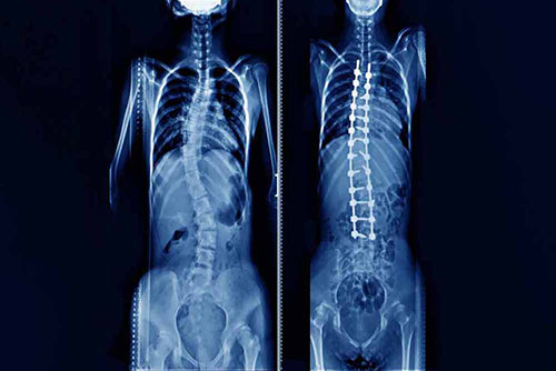
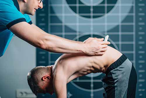
During the actual surgical procedure, the surgeon exposes the affected section of the spine. The operation is performed either from the front, through the chest or abdomen, or from the back. All surgical scoliosis therapies have the common goal of straightening the crooked spine and eliminating its rotation. The doctor also stabilizes the spine, for example using screws and rods.
Therapy by stiffening
With the so-called spondylodesis (spine fusion) one intentionally causes the vertebrae to grow together in the affected area. So you want to stiffen the spine in its previously corrected form.
Recent surgical scoliosis therapies for children and adolescents
A stiffening of the spine prevents its natural growth. Therefore, it is not an option for children and adolescents. Instead, doctors use special titanium rods in these cases, for example.The so-called VEPTRs (vertical expandable prosthetic titanium ribs) are used in such a way that they do not prevent the spine from growing - for example from the rib to the vertebra.
With this scoliosis therapy, the doctors have to regularly adjust the rods to the growth through further minor interventions, approximately every four to six months.
Modern variants of such rods, the "growing rods " (=growing rods), contain a small remote-controlled motor. In this way, they can be adjusted to the respective spinal column growth from the outside and without further intervention.
A complex system of screws, rods and a special plate called the Shilla procedure also promises scoliosis therapy without impeding growth. The rods used "grow" because they slide in their fastening screws. Once the bone growth is complete, the system can be removed again.
Correction system
Another method is the "ApiFix" correction system. It is fixed vertically in the arch of curvature of the scoliosis. Physiotherapeutic treatments follow in the months after the implantation.The correction system reacts to this with a ratchet mechanism: if the spine stretches during an exercise, the system is pulled along and snaps into a new position. As a result, the spine no longer falls back into its original, crooked position. This scoliosis therapy is carried out gradually so that the surrounding tissue adapts better.
Clamp technique
This form of surgical scoliosis therapy is suitable for curvature angles of less than 35 degrees. Doctors attach special, claw-shaped clamps (shape memory alloy, SMA) to the curve of the spine. They are cooled before the procedure, after the procedure they are pushed back into their original shape by the patient's body heat, thus correcting the scoliosis.
Rehabilitation
Depending on the surgical scoliosis therapy carried out, further treatments follow, for example:Scoliosis corset that can be removed once the operated parts of the spine have ossified
Controlled physiotherapeutic applications and physiotherapy exercises
Rehabilitation is either outpatient or inpatient. Those affected are urged to learn new movements as early as possible in any case. With such rehabilitation measures, surgical scoliosis therapy can be sensibly supported and later damage prevented.
Treatment of underlying diseases
If scoliosis is the result of another disease, it is always treated as well. This applies in particular to diseases or malformations that would promote the progression of the spinal curvature. For example, if a patient has legs of different lengths, one tries to compensate for this difference with special shoes.
Pain therapy
Scoliosis pain in the back, neck or shoulders, but also headaches are usually treated with painkillers in tablet form. Patches that emit heat can sometimes help. Local anesthetic injections in the back are only used for severe pain. They arise in the context of scoliosis, for example, due to wear and tear damage to the spine and constricted nerves in the spinal cord.The so-called transcutaneous electrical nerve stimulation ( TENS ) sometimes also helps against pain in scoliosis . Electrodes are placed on the skin over the painful area. These electrodes release electrical impulses that act on deeper nerves. They thus inhibit the transmission of pain from these nerves to the brain. The German Scoliosis Network also lists acupuncture as part of a comprehensive scoliosis therapy - it is also said to relieve pain in some patients.
Scoliosis Exercises
In the case of slight curvatures of the spine, physiotherapy exercises are suitable as scoliosis therapy. You should correct your posture. In addition to physiotherapeutic applications, there are also exercises for scoliosis that patients can carry out at home. Exercise as part of scoliosis therapy should:
Improve posture
Strengthen the muscles
Eliminate bends forward and backward
Increase lung and heart function
There are now many methods of treating scoliosis using exercises.
Read more about how scoliosis can be treated with exercises in the article Scoliosis Exercises .
aids
In addition to a corset, which is used in some cases as a therapy aid, there are other aids for scoliosis patients that may also be reimbursed or subsidized by health insurance companies (depending on the individual case).
For example, there are special pillows and mattresses that help those affected to sleep better or without pain.
In severe cases, walking aids are possible, and special ergonomic office chairs also help those affected in everyday life or at work.
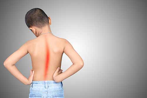
Scoliosis symptoms
In many cases, scoliosis is more of a cosmetic problem. However, the longer it goes untreated, the more likely it is that you will experience pain as the disease progresses. Because how pronounced the symptoms are always depends on how far advanced the curvature is.The external scoliosis symptoms that can be seen with the naked eye include, among others
- Shoulders of different heights
- Basin crooked or protruding on one side
- Tilted head
In the case of pronounced scoliosis, the so-called rib hump often occurs, and in many cases muscle bulges form in the lumbar and neck areas.
Due to increasing signs of wear and tear, those affected have increased problems with muscle tension and pain, especially from the middle of the third decade of life. The lung capacity may also decrease and shortness of breath, a feeling of pressure in the chest or tachycardia may occur in some cases.
Read more about scoliosis symptoms here .
Scoliosis causes and risk factors
About 90 percent of all scoliosis are idiopathic, so you don't know why they develop. In the remaining ten percent - the secondary scoliosis - various causes come into question that lead to a curvature of the spine.
Malformation scoliosis
This form of scoliosis is due to congenital malformations of individual parts of the spine, for example- Wedge-shaped vertebral bodies (different edge heights)
- Split or half formed vertebrae
- Congenital malformations of the ribs (synostoses)
- Defects in the spinal canal (such as diastematomyelia)
Experts therefore refer to it as congenital (congenital) scoliosis.
Myopathic scoliosis
These spinal curvatures are based on muscle diseases (including hereditary muscle weakness diseases). The most common is Duchenne muscular dystrophy, in which a certain muscle protein is not formed. As a result, children suffer from progressive muscle weakness and wasting from an early age. More than half of all those affected develop scoliosis in the course of Duchenne muscular dystrophy, usually in early adolescence and after the loss of the ability to walk.In severe cases, arthrogryposis also often leads to pronounced scoliosis. It is a congenital joint stiffness that is caused by changes in the tendons, muscles and connective tissue.
Neuropathic Skoliosen
In this form, damage to the nervous system leads to a crooked spine. Muscles that stabilize the spine (abdominal and back muscles) then no longer function as usual. This creates an imbalance and causes the spine to curve in the direction of the flaccid muscles.
Among other things, these diseases of the nervous system lead to scoliosis:
- Myasthenia gravis (muscle paralysis)
- Viral spinal cord inflammation (myelitis)
- Early childhood brain damage (such as infantile cerebral palsy)
- Neurodegenerative diseases with damage and loss of nerve cells (e.g. spinal muscular atrophy with loss of the second nerve pathway to the muscles)
- Cavities in the spinal cord due to cerebrospinal fluid congestion (syringomyelia)
- Malignant or benign growths (such as spinal tumors)
Other scoliosis causes
In addition to the muscle and nerve diseases mentioned, numerous other clinical pictures may be associated with scoliosis of varying severity. The surrounding connective tissue, but usually also the bone and cartilage structures are directly affected. The table gives some examples.In addition, accidents sometimes lead to scoliosis. These post-traumatic scoliosis occur, for example, according to the book of a vertebra, burns or spinal cord injuries. Furthermore, some medical procedures cause spinal curvature, such as radiation or laminectomies . In the latter, part of the vertebral bone is removed (vertebral arch, possibly with the spinous process).
As with many diseases, experts suspect that scoliosis is also hereditary. 97 percent have a family history. Up to 70 percent of identical twins will both develop scoliosis. Since scoliosis increases with age, researchers assume that signs of wear and tear ultimately also have a decisive influence (degenerative changes).
Diagnosis of Scoliosis
The orthopaedist is the specialist for diseases of the supporting and musculoskeletal system. There are also pediatricians and pediatric orthopedists who specialize in scoliosis. First, the doctor collects the medical history ( anamnesis ) and asks the patient or the relatives caring for them the following questions, among others: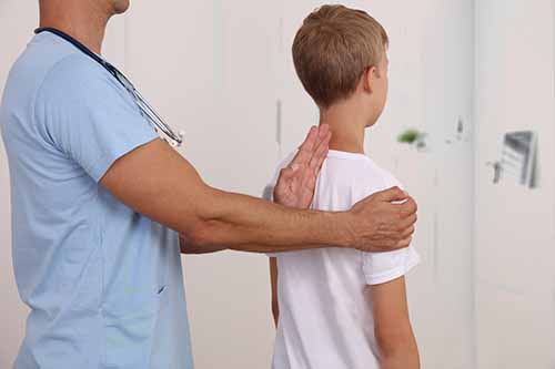
- When did you first notice the crooked spine?
- Do you suffer from complaints such as back pain?
- Has the first menstrual period (menarche) or broken voice already occurred?
- How fast have you grown in recent years?
- Do you have any other known medical conditions, such as foot deformities, a tilted pelvis, muscle or nerve diseases?
- Are there any known cases of scoliosis in your family?
The US Scoliosis Research Society regularly publishes questionnaires for patients suffering from scoliosis (current version SRS-30). In German translation, doctors also use this questionnaire here.
It makes sense for those affected to fill out the questionnaire at regular intervals. This makes it possible to state how they perceive the course of the disease and to assess the success of the therapies carried out.
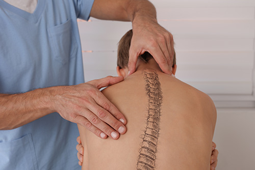
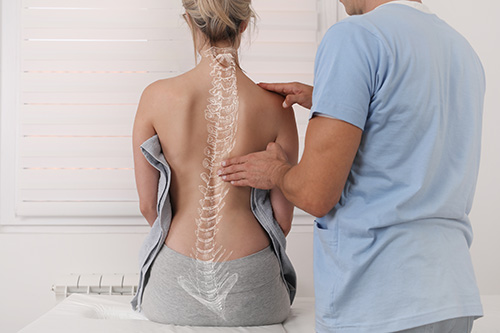
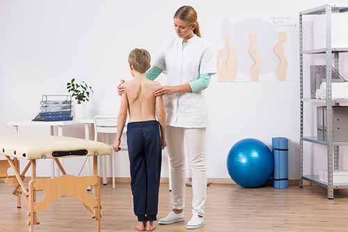
Physical examination
After the questioning, your doctor will examine you physically. First he determines the standing and sitting size, then he examines the back and especially the shape of the spine. If the line of the spinous processes deviates, he determines a so-called overhang. The chest is shifted to the side. As a result, a straight line from the last cervical vertebra down ends in a scoliosis no longer in the anal fold, but next to it.He also checks the lateral equality of the shoulder blades (symmetrical shoulder stand) and the waist as well as the outline of the torso. With scoliosis, the shoulders are different heights. The two so-called waist triangles are also different in size, i.e. the distances from the left or right arm hanging down to the torso.
During the physical examination, the doctor also views the still image from the side. This allows him to detect excessive hunchback (hyperkyphosis) or a spine that is strongly curved towards the abdomen (hyperlordosis, such as a hollow back).
In rare, pronounced cases, a clear thoracic hump forms. The thoracic spine is then not only curved to the side, but also strongly backwards (kypho-scoliosis).
Such kypho-scoliosis usually occurs with other diseases, for example rickets, bone marrow inflammation or tuberculosis of the vertebral bodies.
In addition, a crooked pelvis or legs of different lengths (leg length difference) are also noticeable in the context of scoliosis.
The doctor also examines the skin on the back, as this is where diseases of the spinal cord may already be evident.
Light brown and even spots on the skin, so-called café-au-lait spots, on the other hand, are typical of the hereditary disease neurofibromatosis type 1 (Recklinghausen's disease), which mainly affects the skin and the nervous system. In some cases, those affected also suffer from scoliosis, in particular kypho-scoliosis.
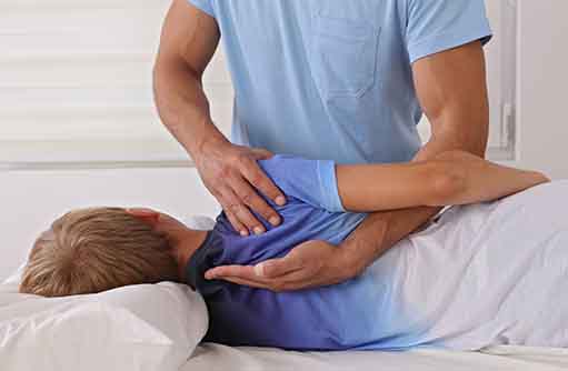
Physical examination of the infant
Scoliosis in infants can be visualized using various holding tests. For example, if the child lies with its stomach on the examiner's hand , he can easily recognize a crooked spine, since the curvature is usually clearly visible on the back.Differences in arm and leg development can be observed in the Vojta side-tilt reaction . To do this, the doctor holds the child sideways and pays attention to the body tension of the infant. When held on the side facing away from the curve, the body usually falls off much more slackly than on the side to which the curve is directed.
Scoliosis is also clearly visible in the vertical hanging reaction according to Peiper and Isbert . Held upside down by the feet, the infant's entire body is curved to one side in a C-shape.
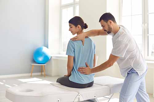
Adams-Test
During this examination, the doctor will ask you to bend forward with your knees straight. If he now looks at her back, he may recognize typical signs of scoliosis, for example a hump on the ribs on the back with a bent posture or muscle bulges in the neck and lumbar area.As a rule, the doctor measures the extent of the rib hump or the muscle bulges using a so-called scoliometer or inclinometer . He compares the heights of the left and the right side with each other. According to the guidelines, deviations of more than five degrees are to be regarded as pathological. In these cases, further examinations follow, in particular x-rays of the spine.
Examination of mobility, strength, flexibility and reflexes
As part of the physical exam, the doctor will also ask you to bend forwards and backwards and to the side. This checks the mobility of the spine. He also measures the finger-to-floor distance in a position bent forward as much as possible with legs stretched out. Ideally you should be touching the floor (0 cm), but this is rarely possible with severe scoliosis.In addition, the doctor will check whether the curvature of the spine can be actively compensated for by your own movements or with the doctor's manual assistance (passive, manual redressability). "Real", structural scoliosis can hardly or not at all be changed.
It is also important to recognize any abnormalities in the nervous system that cause scoliosis or possibly result from a spinal curvature or hereditary connective tissue diseases (Marfan syndrome).
X-ray
In many cases, the doctor diagnoses scoliosis based on the physical examination alone. However, if he suspects a spinal curvature, he will always arrange for an X-ray examination. The entire spine is depicted while standing, viewed from the front (or rear) and from the side.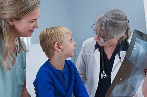
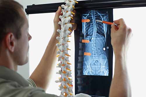
With the help of the X-rays, the doctor measures the Cobb angle (in the case of infant scoliosis, the rib departure angle RVAD), determines the main and secondary curvatures, identifies the vertebrae and the end vertebrae and determines the curvature pattern. This procedure is important for subsequent scoliosis therapy. In addition, malformations or deformations of the bones can be detected.
Determination of skeletal maturity
In order to assess the course of scoliosis in adolescents, it is important to determine the status of spinal growth. To do this, X-ray images are used to assess the skeletal maturity based on the ossification of the iliac crest processes (apophyses).These extensions ossify more and more with increasing age. When they are completely ossified and the apophyses are closed, skeletal growth is complete. Bone age can also be determined using an X-ray of the wrist and classified according to Greulich and Pyle.
Although the age is usually related to the skeletal maturity, it may also differ. Bone age is more reliable than age for the prognosis of scoliosis.
X-ray alternatives
In addition to a conventional X-ray diagnosis, there are also a number of imaging methods without exposure to radiation for examining scoliosis. Alternatives are, for example, the Optimetric method, Moiré photogrammetry, the video grid steriometric Formetric system or the 3D spine analysis "ZEBRIS". Using these methods, however, scoliosis can only be assessed to a limited extent, especially in comparison to X-ray images.
Further investigations
In exceptional cases, the doctor will have sectional images made using a magnetic resonance imaging (MRI) scanner, especially if malformations of the spinal cord or changes in the spinal cord canal (such as tumors) are suspected.In the case of severe scoliosis, the heart and lung functions are disturbed by the curvature and twisting of the entire chest area. In these cases, the doctor arranges further tests. These include, for example, ultrasound examinations of the heart and a lung function test ( spirometry ).
Depending on the extent of the scoliosis, the doctor regularly repeats various tests to monitor the progression of the spinal curvature. In the X-ray examinations for control, however, doctors usually limit themselves to a frontal image.
Course and prognosis of Scoliosis
The course of a scoliosis is very different. In principle, the earlier a spinal curvature occurs, the more likely it is to progress (untreated).An exception is infant scoliosis. Within the first two years of life, a crooked spine recedes on its own in up to 96 percent of cases. It can also be positively influenced by suitable positioning measures and physiotherapy.
If a residual scoliosis of more than 20 degrees remains, the parents of the affected baby must expect the scoliosis to progress.
Risk of worsening scoliosis
If scoliosis only occurs later in life, the prognosis depends on various criteria. For example, underlying diseases of the muscle or nervous system often worsen the course of the disease. In the case of idiopathic scoliosis, other factors are important in addition to age (possible residual growth):
- Initial Cobb angle
- Risser stage (skeletal maturity)
- Time of the first menstrual period (menarche, proven connection with relapsing bone growth in the following years)
The Cobb angle is most important at the beginning of the diagnosis. The guidelines state the likelihood of idiopathic scoliosis increasing, depending on the degree of curvature and age, as follows:
course of the disease in old age
In many cases, scoliosis worsens in adulthood. This is especially true when the Cobb angle is greater than 50 degrees at growth completion. Calculations on thoracic and lumbar scoliosis have shown that the curvature increases by about 0.5 to one degree per year.Severe scoliosis, especially in the lower back, increases the risk of painful symptoms. Particularly pronounced curvatures often also irritate the spinal cord nerves (spinal nerves) and then cause discomfort or pain.
If the scoliosis reaches a value of around 80 degrees, it reduces life expectancy in many cases.
In the case of very pronounced courses, breathing becomes increasingly difficult due to the increasing deformation. The chest is hardly mobile (thoracic rigidity) and the lung volume decreases. On the side of the curve, the lung becomes overinflated (pulmonary emphysema). The other half of the lung, on the other hand, is hardly ventilated and the lung tissue collapses in places ( atelectasis ).
There is a risk of serious complications such as pneumonia, chronic bronchitis or inflammation of the lungs (pleurisy). In addition, the heart is increasingly stressed ( cor pulmonale ).
Complications after scoliosis surgery
Like any surgical procedure, an operation on the spine involves certain risks such as bleeding, infections (especially in acne patients) or wound healing disorders. Sensory disturbances or paralysis do not usually occur in idiopathic scoliosis. However, surgical scoliosis therapy can lead to nerve or spinal cord injuries.However, the probability of such a complication is very low. According to studies, it is between 0.3 and 2.5 percent. The risk increases when a major surgery is performed and other diseases (especially of the spinal cord) are present. In some cases - such as spinal cord disorders - the doctors wake up the person affected during the operation and check their movements and sensations on the skin.
Effusions and "Pneu"
If the operation was performed through the chest cavity, fluid accumulations in the chest are also possible. The doctors guide these out of the body through a tube (drainage). Under certain circumstances, a lung collapses, which is medically referred to as pneumo-thorax (short: pneu). Here, too, a special drainage system is used to allow the lungs to unfold again.
correction loss
After some stiffening operations, the counter-curvature of a scoliosis also increases. In addition, the correction achieved is sometimes partially lost in the first years after the procedure. However, scoliosis usually stabilizes after the operation.In young patients who are stiffened at an early age (Risser 0), a loss of correction can be problematic. As the vertebral bodies continue to grow, spinal torsion increases in many cases. Doctors speak of the so-called crankshaft phenomenon . To prevent this, stiffening scoliosis therapy is usually performed from both the front and the back.
Other specific complications include metal fractures of the rods and screws used during an operation. In these cases there is almost always a loss of correction. In some stiffening operations, the vertebral bodies do not grow together as planned. "Wrong" joints form, so-called pseudarthroses. They may cause persistent pain (particularly in lumbar scoliosis).
Some patients develop a flat back as a result of straightening surgery using rods (Harrington rods). The naturally existing forward curvature of the lumbar spine is eliminated. As a result, the adjacent vertebrae and discs wear out faster, causing painful discomfort.
Scoliosis and pregnancy
Contrary to many fears, scoliosis does not have a negative effect on pregnancy. It does not matter whether the patients were treated conservatively (physiotherapy, corset) or surgically. As with all pregnant women, those affected by scoliosis sometimes experience lower back pain, but an increase in the Cobb angle has not yet been demonstrated.
Control examinations
Depending on the extent of the scoliosis, the doctor regularly checks the course of the curvature. Children with spine curvatures less than 20 degrees are checked by physical exams about every three to six months. If the doctor suspects an increase in curvature, he will order an X-ray. Scoliosis over 20 degrees is checked at least once a year by means of an X-ray examination. Clinical examinations are also carried out at least every six months as part of scoliosis therapy.Once growth is complete and the Cobb angle is less than 20 degrees, no further control is required. In the case of a scoliosis of 20 to 40 degrees that has not been operated on, the doctor carries out a check-up about two to four years after growth has ended. If the curvature has increased by five degrees, further checks follow. If adults suffer from scoliosis over 40 degrees, the guidelines recommend an annual review.
If the affected person has had an operation, no further routine examinations are necessary two years after the operation if the fusion is stable and the Cobb angle is less than 40 degrees.
Living with scoliosis
In most cases, patients live well with their scoliosis. It is important to work actively against the spinal deformity. Integrate scoliosis exercises into your everyday life.Do (school) sports. Various sports are suitable for this, such as various forms of yoga, swimming — especially backstroke. Archery, cycling, Nordic walking or therapeutic horseback riding are also considered suitable sports. If you have concerns about some activities, be sure to consult your doctor.
If your scoliosis burdens you in everyday life, for example at work or in your free time, do not hesitate to ask for help. Contact your employer, your physiotherapist or friends. Some of those affected are also involved in self-help groups.
If the scoliosis weighs heavily on your psyche or that of your child, psychotherapeutic treatment may also make sense. By being open and active, you will collect valuable tips and prevent the progression of your scoliosis .
Prevention
Since the causes of most scoliosis are not known, scoliosis cannot generally be prevented. In the case of known high-risk diseases, however, it helps to identify the onset of scoliosis in good time through regular check-ups and to prevent it from getting worse.
The same applies to the standard check-ups for children and adolescents, with which a diagnosis can be made early in the growth phase. Appropriate therapy can prevent scoliosis from progressing and subsequent consequential damage.
There are no comments yet. Would you like to add a comment?
In accordance with Article 10 of the Personal Data Protection Law (PDPL,KVKK) titled Data Controller's Obligation to Disclose, we use cookies in accordance with the legislation, limited to the purposes specified in the privacy policy.