Cataract Surgery in Turkey, Prices, Procedure and Aftercare
What is a cataract?
With a cataract , the lens of the eye gradually becomes cloudy. Often as a result of natural aging. The only effective treatment option is surgery . The cloudy lens is removed and replaced with a new, artificial lens .
Causes of cataracts
The following causes can lead to a clouding of the eye lens and thus to a so-called cataract :
- aging of the lens of the eye
- Metabolic disorders of the lens of the eye
- Trauma (e.g. hit to the eye)
- Congenital opacities
The age-related cataract
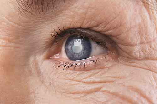
Age-related cataracts are the most common form of opacity in the lens of the eye. The aging process of the lens of the eye begins in many patients from the age of 40 and initially manifests itself as presbyopia or presbyopia.More than 80% of Europeans have a measurable cataract from the age of 60. However, not every clouding of the lens has to be accompanied by a visual impairment. However, if the clouding of the lens already leads to a visual impairment, then one speaks of a cataract.
The definition of when a cataract is present depends only on the visual function and not on the actual cloudiness of the lens.
Most statutory health and health insurance companies only recognize a cataract diagnosis if the visual impairment that has occurred means that driving a motor vehicle is no longer possible or at risk and only then will they cover the costs of the cataract operation.
Metabolic disorders as a cause of cataracts
Metabolic diseases - above all diabetes - can significantly accelerate clouding of the eye lens: Large fluctuations in blood sugar levels can lead to water retention in the eye lens, which can lead to clouding and thickening of the lens.Drugs can also be the cause of an earlier onset of a cataract: Long-term use of cortisone inhalants for asthma or chemotherapy can lead to a metabolic disorder in the lens of the eye and thus to cataracts.
Accidents and trauma as a cataract cause
A blow to the eye can cause clouding of the lens of the eye, which can lead to cataracts even decades after the actual trauma.This form of mostly unilateral cataracts often shows a typical star-shaped opacity of the nucleus of the eye lens. This typically leads to very high glare sensitivity and night blindness.
Congenital cataracts
In addition to childhood cataracts, which are usually operated on before the child is five years old, there are also milder forms of congenital cataracts that only affect parts of the lens of the eye. These little “islands of cloudiness” can go unnoticed for years. However, they can increase with age and lead to poor eyesight.
Cataract Symptoms
The signs and symptoms of cataracts are
- Increased need for light, especially when reading
- Increased sensitivity to glare at night when driving or night blindness
- One-eyed double vision
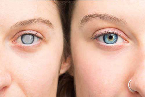
- Gray haze or color weakness
- "Gray Pupil"
- Exhausted, watery eyes
- Visual impairment that cannot be corrected with glasses
- Increased eye pressure or glaucoma
When glasses no longer help
The cataract leads to a change in the lenses: the values in the glasses can change again and again within months. In the beginning it is often still possible to correct the visual impairment with new glasses.With increasing cloudiness, the visual impairment can no longer be corrected with new glasses.
Here, cataract surgery is the most sustainable solution to ensure stable eyesight and improved visual performance.
For this purpose, an artificial lens is inserted into the eye in a surgical procedure.
Depending on the choice of lens implant and the suitability of the eyes, lens implants can be chosen for cataract surgery that make glasses superfluous.
High-quality multifocal lenses are able to permanently correct almost all forms of nearsightedness, farsightedness, astigmatism and presbyopia.
Cataract Diagnosis
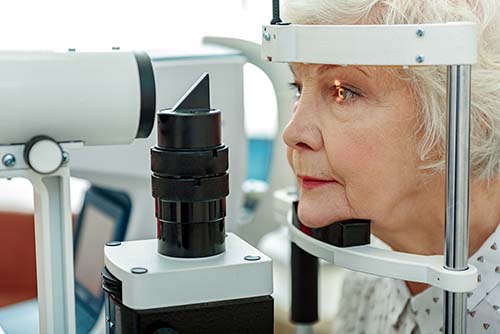
Before Cataract Surgery - Diagnosing cataracts involves examining visual function, the anterior segment of the eye, and ruling out other causes of vision loss.
Measurement of eyesight and visual performance
Digital autorefractometers allow an exact measurement of visual acuity and visual performance. The collected values are checked by the ophthalmologist using subjective refraction.The autorefractometer is used to automatically determine the objective refraction (visual acuity test).
Examination with the slit lamp (biomicroscopy)
Here the cornea, iris and lens are examined. This examination can already detect advanced clouding of the lens of the eye.Cataract examination by slit lamp
Corneal analysis (pentacam analysis)
The entire front section of the eye is recorded using a three-dimensional scan: Here, precise measurement of lens opacity and analysis of abnormalities on the cornea and astigmatism is possible.Endothelial analysis of the cornea (endothelial microscopy)
This technique makes it possible to examine the "cell density" of the cornea. The information obtained is very important for choosing the right lens implant for cataract surgery. A low or conspicuous cell density of the cornea can, for example, be an important reason for preventing the use of multifocal lenses.Cataract preliminary examination - endothelial analysis with the Pentacam
Examination of the retina (Optomap Scan)
Using this laser-controlled scan, the entire area of the seeing retina as well as the visual center and the optic nerve can be displayed.Abnormalities that lead to a reduction in vision can be discovered here. This information is also very important for choosing the right lens implant.
Layer image of the macula (OCT scan)
This non-contact recording technique allows an analysis of the retinal cell layers in the area of the visual center. Abnormalities here can also have an impact on the correct choice of a suitable implantable lens.
Laser biometrics
This procedure allows the lens power to be determined for the lens implant. With the right lens strength, an existing ametropia can be corrected at the same time as the operation, so that after the operation, for example, glasses are no longer required (depending on the lens implant).
Cataract treatment
Cataracts are not actually a disease. Rather, it is the result of the natural aging process of the lens of the eye . This hardens over the years and becomes cloudy. Hardening leads to presbyopia ( presbyopia ) , clouding of the lens to cataracts. In general, one speaks of cataracts as soon as visual performance decreases and safe driving ability is no longer guaranteed. To treat cataracts , the clouded endogenous lenses are removed and artificial lenses are implanted . Depending on which lens is used, visual defects can be permanently corrected and freedom from glasses can be achieved for life .
In order to correct presbyopia, the lenses can also be replaced before they become cloudy. Every year, more and more people decide to have multifocal lenses implanted in order to no longer be dependent on visual aids such as contact lenses or progressive lenses.
Examinations before cataract surgery
The decisive factor is often the examination of the visual acuity and the current strength of the glasses . If full vision can no longer be achieved despite glasses correction, this is often an indication of lens clouding. A precise examination of the lens by the ophthalmologist then usually brings certainty. The cloudiness of the lens can also be measured using special diagnostic devices – so-called Scheimpflug cameras.Before an operation can be performed, a decision must be made on a lens implant. This requires a very precise examination of the retina and cornea. The basis for lens calculation is optical biometry and the corneal topography and the examination of the corneal endothelium are particularly important for the correct choice of lens .
Because diseases of the corneal endothelium can be an exclusion criterion for the implantation of multifocal lenses. The condition of the corneal endothelium also plays an important role in the choice of surgical procedure. The examination with the slit lamp and the control of the intraocular pressure serve to rule out eye diseases.
Surgical procedure for removing the lens in cataracts
Ultrasonic procedure (phacoemulsification)
This procedure is the standard procedure for lens removal . First, the eye surgeon makes 2-3 very small incisions (1 to 3 mm) to get inside the eye. A vibrating hollow probe is then inserted into the lens capsule. The ultrasonic energy breaks up the lens material and sucks out the small fragments . This process has been tried and tested for more than 30 years and is inexpensive.
Handling the hollow probe requires a great deal of experience on the part of the operator . This is important so that the spherically emitted ultrasound energy does not damage the cornea or the lens capsule. The harder the lens, the hotter the probe can get. Therefore, sufficient cooling of the probe must be ensured during the operation. The ultrasound procedure is accepted by all statutory and private health insurers.
Femtolaser method (FemtoCat)
In this more recent procedure, the operation is facilitated by a cutting femtosecond laser. The femtolaser prepares the cuts on the cornea and opens the lens capsule . By making cuts in the lens, the laser can prepare the division of the lens nucleus. The surgeon then makes the actual cuts and suctioning out the lens fragments manually. He can use the classic ultrasonic hollow probe to remove the lens. The femtolaser process is very promising for the future. However , the medical advantage of the procedure is currently still very controversial. Therefore, this procedure is usually not covered by health insurance companies. In the case of private insurance companies, it remains a case-by-case decision. The additional costs are 1000 to 1500 euros per eye.
Nanolaser (Photo-Phaco-Disruption)
nanolaser probe. An important hygienic advantage of the nanolaser: the lens removal is only carried out with disposable probes and reduces the risk of infection.An important hygienic advantage of the nanolaser: the lens removal is only carried out with disposable probes and reduces the risk of infection.
This modern method replaces the classic ultrasonic hollow probe. A very fine laser probe enables the actual lens removal here. Laser pulses in the nanosecond range are emitted within the hollow probe. The laser energy breaks up the lens contents and the probe sucks them out.
No heat energy is generated by the laser and the risk of injury to the cornea or lens capsule is significantly lower . That is why the use of the nanolaser is a good choice, especially for corneal diseases.
The laser probe is a disposable item . A disinfection and preparation of the probe for use by the next patient therefore does not take place here. This is an important hygienic advantage of the nanolaser process compared to the classic ultrasound process.
Due to the lower energy release in the eye and the higher hygienic safety, the nanolaser procedure is already covered by some private health insurance companies for diseases of the cornea. Statutory health insurance companies cannot cover the additional costs. The additional costs are 1000 to 1500 euros per eye. In Munich, the nanolaser process is currently only available in our eye center .
Anesthesia
Cataract surgery takes an average of 10 to 20 minutes per eye. Experienced eye surgeons usually perform the procedure under topical anesthesia and short twilight sleep . As a standard procedure, some surgeons use an anesthetic injection on the eye to immobilize the entire surgical area. General anesthesia is possible, but usually only necessary for children.A comparison of implantable lenses: lens selection for cataract surgery
Once the lens of the eye has been removed, the new lens can be inserted into the existing lens capsule. Modern lenses are soft and foldable and can be inserted into the eye through a 2mm port. They unfold in the eye and are inserted into the lens capsule behind the pupil - invisible from the outside . Today there is a wide range of lens implants:
Standard lenses
These lenses have a fixed focal length. Unfortunately, correction of astigmatism is not possible with standard lenses. Likewise, no light protection filters (e.g. violet/blue light) are installed here. After an operation with standard lenses, a reading aid or varifocal glasses and the wearing of light protection glasses are usually required.CT Aspina: The standard lens has a fixed focal length.
The standard lens has a fixed focal length.
Premium lenses
These lenses also have a fixed focal length. However, they are optimized for night vision (aspherical) and/or equipped with a protective filter and/or a contrast-enhancing filter. Here, too, a reading aid or varifocal glasses is usually required to cover all visual areas.Premium lens is optimized for night vision and equipped with a protective filter or a contrast-enhancing filter.
The premium lens is optimized for night vision and equipped with a protective filter or a contrast-enhancing filter.
Toric lenses
These lenses belong to the premium lenses. These can also correct astigmatism and thus generally make the distance or near range glasses-free.The toric lens enables reliable astigmatism correction.
The toric lens enables reliable astigmatism correction.
Multifocal lenses
Multifocal lenses usually have light protection filters and can also correct astigmatism. The special feature is that these lenses can cover both the long and close range . With modern multifocal lenses, a 95% freedom from glasses can be achieved in addition to the protective functions.However, a prerequisite for successfully going without glasses is good suitability: no irregular astigmatism, normal corneal endothelium and adequate three-dimensional vision. These criteria should definitely be checked before recommending a multifocal lens.

The additional costs for premium and multifocal lenses are 400 to 1,600 euros per lens.
There are no comments yet. Would you like to add a comment?